Need Assistance? Call us today! 602-478-9713
Dr Dave Singh DMD PhD DDSc © 2024
Adjunct Professor, Sleep Medicine, Stanford University, USA
Idea/Innovation
I am often asked how I came up with the idea of a biomimetic device, such as the DNA appliance, based on the concept of craniofacial epigenetics for the treatment of obstructive sleep apnea. The story begins in 2001 at the Center for Craniofacial Disorders, University of Puerto Rico, USA where a team of dedicated clinicians were developing techniques to treat babies, children and adolescents with a history of craniofacial congenital abnormalities, ranging from cleft lip and palate through to craniosynostoses, including Apert syndrome, etc. At that time, it was decided that these pediatric patients would be treated with a new technique called Distraction Osteogenesis. The UPR Center for Craniofacial Disorders was one of the first centers in the world to investigate this novel technique for midfacial advancement in these types of cases. One of my roles in this team was to undertake quantitative assessment of morphologic changes of the skeletal and soft tissues pre- and post-treatment. At the turn of the century, cone-beam computerized tomography (CBCT) technology was not available, so we used non-ionizing magnetic resonance imaging (MRI) scans and I introduced 3D stereophotogrammetry for surface imaging of these babies. To get a rigorous determination of shape, size and directional changes, I utilized a new series of 3D techniques, including geometric morphometrics for mathematical modeling, which had never been deployed before. These elegant techniques permit determination of changes after correcting for size differences in statistical shape-space, and the results can be pseudo-colored to permit intuitive interpretation of the outcomes by clinicians.
At the heart of innovation lies observation. I noticed that nearly all of the cases that were being considered for surgical correction had midfacial hypoplasia prior to treatment. Some of these cases had a genetic mutation such as Apert syndrome where the facial sutures had undergone premature synostosis, and the midface was unable to develop normally because of this. Using the distraction osteogenesis protocol, the craniofacial and plastic surgeons carefully induced a controlled osteotomy. Next, a rigid external distractor was placed, and the patient underwent latency, allowing wound healing to occur. This was then followed by distraction of the callus whereby the orthodontist would manipulate and advance the midface, using the external distractor screws. After approximately 6 weeks, the patients entered the fourth phase, consolidation, to allow the tissues to heal in the new position, after which the distractor was removed. After this procedure, I noticed that not only did the patients have an improved appearance, but their demeanor also improved, and that intrigued me. I went back and analyzed the MRI data. To my surprise, the upper airway had ballooned in all of these cases, and I surmised that these children were therefore sleeping better because of that, which led to daytime improvements in their behavior. Recalling that these children had craniosynostoses, and that the general population does not have this genetic mutation, I wondered if a similar non-surgical technique could be developed for more generalized use to address upper airway issues, specifically obstructive sleep apnea.
 Current competitors
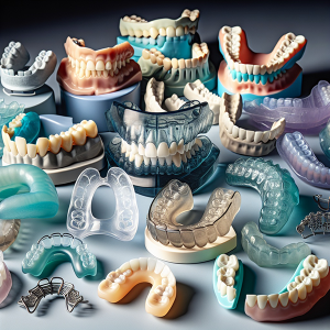
Before launching into a futile effort, I decided to undertake a comprehensive review of as many orthodontic devices that I could find that had historically or were currently being used for maxillary or palatal expansion. These devices included rapid palatal expanders, functional appliances and others that were used by orthodontists and general dentists for these types of purposes. In all, I reviewed nearly 250 appliances, and several of my formal studies were published in the peer-reviewed medical, dental and orthodontic literature. I noticed that these techniques were based on what some might say are ‘outdated’ principles. For example, rapid palatal expanders were used to split the midpalatal suture transversely, induce a midline diastema and then the teeth were moved orthodontically to close the gap. Even more controversial was the idea that palatal expansion could be undertaken in adults where the sutures had undergone “fusion”.
In addition, when ‘jumping the bite’ (mandibular advancement using a ‘functional appliance’) for Class II orthodontic correction, there was, and still is, controversy as to whether the mandible simply moves forward, actually grows into the new forward position, or if the clinical outcome is some combination of the two. My approach was to ‘allow the data to speak’. In other words, I based my decision on raw clinical data, initially collected from orthodontists in the US, and also from general dentists that were experienced in orthodontics. I reached the conclusion that while each device or technique had certain characteristics or advantages, I could not find an appliance that addressed all four craniofacial tissues: hard tissues (bone); soft tissues (mostly muscle); dental tissues (teeth), and functional spaces (the upper airway) for non-surgical, craniofacial correction.
Background, literature review and mechanisms
Before thinking about device design, it was important for me to research and understand the mechanism(s) through which these appliances putatively worked. My experience in molecular biology/molecular genetics came into play. At that time, stem cells were in the news but there were little or no known applications in the dental/orthodontic space. In addition, the human genome was sequenced in 2003, and various pieces of the biologic jigsaw were being put into place. Around that time also, functional genomics was being investigated and the idea that genes undergo environmental interactions or epigenetics to produce the final clinical phenotype was gaining momentum. Add to this mix temporo-spatial patterning, which suggests that certain genes are expressed at certain times during development to form the body plan, which includes 32 teeth; and the stage was set for further innovation, research and development. But what cohesive theory or hypothesis could provide an explanation for the prediction of clinical outcomes? The older theories were steeped in Newtonian physics and Darwinian genetics. Because of this deficiency, I wrote the Spatial Matrix Hypothesis, published in the University of Michigan Craniofacial Growth Series, which appears to have withstood the test of time thus far. The encompassing concept emerged as Biomimetics. Restorative dentists were already using biomimetic dental materials that mimicked the behavior of dental tissues. The next step was to design a biomimetic device that would mimic natural craniofacial growth and development.
Intellectual property, device design and clinical protocol
Clinical data dictated the design, materials and protocols for the new device. However, to protect the intellectual property, I submitted several US, Canadian and European patents, all of which were eventually issued. One of the first patented components was a unique orthodontic spring design. I had noted that all prior orthodontic springs made point contact with the teeth and acted as finger springs that simply tipped the teeth. My design differed significantly in that it was the first, compressible 3D orthodontic spring with a large surface area. This property was crucial since the nature of the super-elastic, nickel-free wire meant that an intermittent cyclic signal could be imparted to the periodontium through the crown of the tooth to signal stem cells in the periodontium. Next, the appliance had a midline expansive mechanism such as a jack screw or omega loop, unlike preformed templates. This design component permits activation of the midpalatal (and other) suture(s). Moreover, my published clinical studies suggested that concentric collapse of the maxilla occurs in adults diagnosed with obstructive sleep apnea (OSA). This finding had never been reported before and, therefore, a Y-split design with a 3-way screw system was preferential for clinical correction.

Dr. Eugene Azuma, Dr. Dave Singh, Dr. Jerry Hu, Dr. Cecile Sebastian at the ASBA's Sleep & Wellness Conference April 2024
Source: Dr Dave Singh DMD PhD DDSc © 2024
Adjunct Professor, Sleep Medicine, Stanford University, USA